What Is Glaucoma?
Glaucoma is an eye condition where damage to the optic nerve results in gradual vision loss. The optic nerve, the eye's 'electrical cable', carries images from the retina to the brain; hence, any damage to the nerve results in damage to sight.
What Causes Glaucoma?
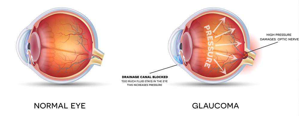
The most common cause of glaucoma is an increase in pressure in the eye. The pressure presses on the nerve, damaging it. Pressure in the eye is needed to help keep its shape and one’s vision in focus. Normal eye pressure is maintained by a process where nourishing fluid is produced by the eye and is then drained out via a meshwork at the back of the eye. When this drainage channel clogs up and fluid begins to build up, high eye pressure occurs. The increased pressure in the eyeball pushes on the optic nerve, damaging it slowly and irreversibly. Glaucoma is one of the world's leading causes of blindness. While it is not curable, blindness can be controlled if it is diagnosed and treated early. Although there are few warning symptoms for glaucoma, regular eye tests can help detect the onset of the disease.
What Are the Different Types of Glaucoma?
There are two main types of glaucoma:
Primary Open-Angle Glaucoma (POAG)
This is the most common form of glaucoma worldwide. It usually affects both eyes and develops slowly such that loss of sight is gradual. Glaucoma symptoms can be subtle or absent, so you may not experience acute eye pain, redness, or significant changes in vision.
Primary Angle-Closure Glaucoma (PACG)
- Acute Angle-Closure Glaucoma
This is a common form of glaucoma in Singapore and occurs more commonly in middle-aged or elderly Chinese females, especially those who are long-sighted. It is characterised by a sudden increase in eyeball pressure. This is due to a blockage preventing fluid from flowing out of the eye. There is a high risk of damage to the optic nerve and so treatment to lower the pressure is urgent to save vision.
You may have Acute Angle-Closure Glaucoma if you experience a sudden onset of pain, redness and blurred vision in one or both eyes. There can also be mistiness of vision and haloes may appear around lights. The pain can be very severe and is often accompanied by headache, nausea and vomiting.
- Chronic Angle-Closure Glaucoma
If left untreated, the condition can rapidly progress to blindness. It is usually caused by a gradual rise in pressure in the eye. Its slow and silent development over a prolonged period means there are no noticeable symptoms except for a progressive vision loss. The disease is often diagnosed at the advanced stage before visual loss is noted. Side (peripheral) vision is usually affected first, whilst central vision remains unaffected. Sometimes, other diseases of the eye can cause a rise in the pressure within the eye; these are classified as secondary glaucoma. There is also a congenital form of the disease present from birth known as congenital glaucoma.
What Are the Symptoms of Glaucoma?
In open-angle glaucoma and chronic angle-closure glaucoma, you may not notice any vision loss until the advanced stages. Vision loss is gradual, with side vision typically affected first. As the condition progresses, the visual field becomes increasingly narrowed. Eventually, only a small central island of vision remains, and you will only be able to see what is directly ahead of you.
In acute angle-closure glaucoma, symptoms are typically more sudden and severe. You may notice the following:
- Blurred vision
- Eye redness
- Eye pain, often associated with headache
- Nausea and vomiting
Who Is at Risk of Getting Glaucoma?
You have a greater risk of glaucoma if:
- A member of your family, particularly a first degree relative, has been diagnosed with glaucoma.
- You have severe myopia (short-sightedness). It is also a natural aging change that occurs in many people simply as a result of growing older.
How Is Glaucoma Detected?
To determine if you have glaucoma, you will be required to undergo a series of pain-free tests.
* Some of these will involve eyedrops that may temporarily blur your vision. You should therefore avoid driving on the day of your appointment.
You may also undergo one of the following:
The ophthalmologist holds a special contact lens against the eye. This allows them to see the area where the fluid in the eye normally drains and check for blockage or narrowing.
Visual Field Test
This measures the range of your 'all around' vision to check for any visual loss. The test takes about 5 minutes per eye. You will be asked to sit at a screen and fix your gaze on a central light target. A series of other lights are then shone, and you will need to press a button indicating if you can see them. This will detect any missing areas in the visual field caused by damage to the optic nerve.
Optic Nerve Assessment
Eyedrops will be placed in your eyes to make your pupils larger. Your doctor will then look at the back of the eye and check your optic nerve. The doctor may take photographs of your eye or use computerised imaging.
How Will Glaucoma Affect Me?
Treatment can stabilise the disease and with regular check-ups, you will be able to manage the condition. You will experience some degree of vision loss but it will be minimal with effective treatment. You will not go completely blind especially if you have been diagnosed at an early stage.
What Are Some Treatments for Glaucoma?
Glaucoma can be treated but not cured. Any vision loss you might already have will be permanent. The aim of treatment is to lower the pressure in the eye and prevent any further damage from developing. It is also important that treatment be maintained throughout your life as it only controls the condition, rather than cure it. It is therefore essential that you comply with the course of treatment given to you as not doing so may result in further loss of sight.
Eyedrops
These are often prescribed in the early stages of glaucoma to lower the eye pressure. Some of the eyedrops may have side effects, usually as a result of being absorbed too quickly into the bloodstream. You can control this by pressing a finger against the lower eyelid where it meets your nose or closing your eye for a minute.
You should consult your doctor if you experience breathlessness or any other side effects.
Laser
Lasers can be used for angle closure glaucoma to widen the angle. In other forms of glaucoma, lasers can also be used to lower the pressure on the eye.
Two types of lasers are applied into the iris. The first helps to create an opening that allows the fluid to flow from the back to the front chamber of the eye, which has the drainage channels. The second laser stretches the iris and opens the angle of the eye. This will enhance the outflow of fluid and lower the pressure. Complications are very rare and include slight inflammation or mild spikes of eye pressure.
Micropulse cyclophotocoagulation is another type of laser that is used to lower the production of fluid within the eye, which helps to temporarily control the pressure.
Glaucoma Surgery
For more severe cases of glaucoma, surgery may be necessary.
Minimally invasive glaucoma surgery (MIGS) can be performed, sometimes together with cataract surgery to help the outflow of fluid through the natural angle.
Another common operation is a trabeculectomy, where a small piece of tissue is removed to allow for a new pathway to drain fluid from the eye. If the trabeculectomy fails to bring the pressure down adequately, a tube insertion under the conjunctiva is required to facilitate fluid outflow. Complications are mostly non-threatening and reversible, and typically resolve spontaneously; these may include ocular irritation, infection and double vision.
Post-operative wound care is very important because it will determine the success of fluid flow. Eye drops that contain anti-inflammatory agents are to be used properly to prevent early wound healing that can cause drainage failure. Anti-infective eyedrops should be used for a shorter time to prevent infection.